


Cowberry Exobasidium (Exobasidium vaccinii)
- Diviziona: Basidiomycota (Basidiomycetes)
- Subdivision: Ustilaginomycotina ()
- Class: Exobasidiomycetes (Exobazidiomycetes)
- Подкласс: Exobasidiomycetidae
- Order: Exobasidiales (Exobasidial)
- Family: Exobasidiaceae (Exobasidiaceae)
- Genus: Exobasidium (Exobasidium)
- Type: Exobasidium vaccinii (Cowberry Exobasidium)
 Mihanaka:
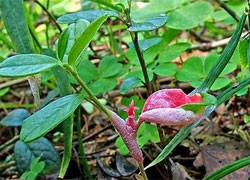
Exobasidium lingonberry (Exobasidium vaccinii) is found very often in almost all taiga forests up to the northern border of the forest in the Arctic. At the beginning or middle of summer, the leaves, and sometimes the young stalks of lingonberries, are deformed: the infected areas of the leaves grow, the surface of the area on the upper side of the leaves becomes concave and becomes red in color. On the underside of the leaves, the affected areas are convex, snow-white. The deformed area becomes thicker (3-10 times in comparison with normal leaves). Sometimes the stems are deformed: they thicken, bend and turn white. Occasionally, flowers are also affected. Under the microscope, it is easy to establish large changes in the structure of the leaf tissue. Cells are noticeably larger than normal sizes (hypertrophy), they are larger than normal. Chlorophyll is absent in the cells of the affected areas, but a red pigment, anthocyanin, appears in the cell sap. It gives the affected leaves a red color.
The hyphae of the fungus are visible between the cells of the lingonberry, there are more of them near the lower surface of the leaf. Thicker hyphae grow between the epidermal cells; on them, under the cuticle, young basidia develop. The cuticle is torn, shed in pieces, and on each mature basidium 2-6 spindle-shaped basidiospores are formed. From them, a gentle, frost-like white coating, noticeable on the underside of the affected leaf, appears. Basidiospores, falling into a drop of water, soon become 3-5-celled. From both ends, the spores grow along a thin hypha, from the ends of which small conidia are laced. They can, in turn, form blastospores. Otherwise, those basidiospores germinate that fall on young lingonberry leaves. The hyphae that arise during germination penetrate through the stomata of the leaves into the plant, and mycelium is formed there. After 4-5 days, yellowish spots appear on the leaves, and after another week, the lingonberry disease has a typical picture. Basidium is formed, new spores are released.
The full development cycle of Exobasidium lingonberry (Exobasidium vaccinii) requires less than two weeks. Exobasidium lingonberry (Exobasidium vaccinii) is the object and cause of controversy for many generations of mycologists. Some scientists see exobasidial fungi as a primitive group, which confirms the hypothesis of the origin of hymenomycetes from parasitic fungi; therefore, these fungi are represented in their systems in an independent order ahead of all other hymenomycetes. Others, like the author of these lines, consider exobasidial fungi as a highly specialized group of fungi, as a side branch of the development of saprotrophic primitive hymenomycetes.
Description:
The fruiting body of Exobasidium lingonberry (Exobasidium vaccinii) is absent. First, 5-7 days after infection, yellow-brown spots appear on top of the leaves, which turn red after a week. The spot occupies part of the leaf or almost the entire leaf, from above it is pressed into the deformed leaf with a depth of 0,2-0,3 cm and a size of 0,5-0,8 cm, crimson red (anthocyanin). At the bottom of the leaf there is a thickened bulge, a tumor-like growth 0,4-0,5 cm in size, with an uneven surface and with a white coating (basidiospores).
pulp:
Ny fitoviana:
With other specialized species of Exobasidium: on blueberries (Exobasidium myrtilli), cranberries, bearberries and other heathers.
Tombana: